| Attribut | Détails |
|---|---|
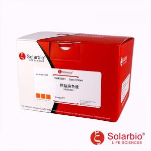
| Chat | G3282 |
| Taille | 2 x 50 ml / 2 x 100 ml |
| Stockage | RT, éviter la lumière, valable 1 année |
Introduction
- Hemosiderin Characteristics:
- Derived from hemoglobin breakdown, it appears golden-yellow or brownish-yellow due to iron content.
- Formed when macrophages engulf red blood cells and break down hemoglobin into iron-free orange blood and iron-containing hemosiderin.
- Perls Prussian Blue Reaction:
- Also known as hemosiderin staining.
- Produces a blue color when treated with potassium ferrocyanide and dilute acid.
- Commonly seen in phagocytes’ interstitium, displaying ferric iron salts.
- Classical histochemical reaction, sensitive, and excellent for displaying ferric iron in tissues.
- The principle involves separating ferric iron from protein using potassium ferrocyanide solution and hydrochloric acid, forming an insoluble Prussian blue compound.
- Stable ferrocyanide of ferric iron allows re-dyeing with red dye after reaction.
- Perls Stain Usage:
- Commonly used to display hemorrhagic lesions, especially in phagocytes.
- Helps determine hemosiderin deposition and distinguish it from other pigments.
- The staining solution is stable, long-lasting without precipitation, and suitable for a wide range of applications.
- Can be re-dyed, making it versatile for different staining techniques.
Composants du kit
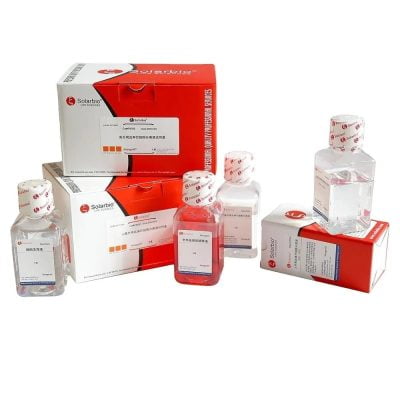
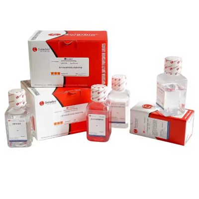
| Réactif | Volume | Stockage |
|---|---|---|
| Réactif(UN): Perls Stain | A1: 25ml <br> A-2: 25ml | 50ml <br> RT, éviter la lumière |
| Réactif(B): Neutral Red Solution | 50ml <br> 100ml | RT, éviter la lumière |
Avant utilisation, mix equal parts of A1 and A-2 to form Perls Stain. It is not suitable to prepare in advance.
Matériaux autonomes
10% neutral formalin fixative, Series of ethanol, Eau distillée, 4% paraformaldéhyde
Protocole (pour référence seulement)
(1) For paraffin section staining
- Fixez le tissu dans 10% natural formalin fixative, dehydrate, and embed.
- Cut sections 4µm thick, dewax to distilled water, and rinse for 1 minute.
- Soak the section in Perls Stain for 15-30 minutes. Rinse fully in distilled water for 2-5 minutes.
- Lightly stain the nucleus with a Neutral Red Solution for 15-30 secondes. Rinse in tap water for 1-5 secondes.
- Déshydrater conventionnellement, transparent, et sceller avec du résinène.
(2) For frozen section staining
- Without dewaxing, rinse directly and quickly with distilled water for 2-3 minutes.
- Follow the other steps as paraffin sections.
(3) For cultured cell staining
- Fix in 4% paraformaldéhyde pour 10-20 minutes.
- Rinse in tap water twice for 2 minutes à chaque fois.
- The steps of staining, déshydratation, transparency, and sealing are the same as paraffin section steps. Adjust time accordingly.
Résultat
- Hemosiderin or Ferric Iron: Bleu
- Nucleus and other Tissues: Rouge
Contrôle négatif
Take the same adjacent section, dewax to water. After incubation in 5% oxalic acid for 2-6 heures, follow the same procedure as above. The result should be negative.
Note
- Ensure clean section dewaxing.
- Utiliser 10% neutral formalin for tissue fixation. Long-term fixation with common formalin can damage tissues. Avoid acid fixatives, as chromate treatment hinders iron preservation.
- Keep containers clean and avoid using metal iron products. Use distilled water for washing sections and containers to prevent iron contamination.
- Adjust Perls Stain dyeing time according to the sample.
- Use the same positive control section for all sections. Autopsy lung tissue is a good control, containing many iron-positive macrophages (heart failure cells).
- Replace ethanol series frequently.
- For frozen section and cell staining, explore experimental conditions based on specific conditions. Wear experimental clothes and disposable gloves for health and safety.
Commentaires
Il n'y a pas encore de critiques.