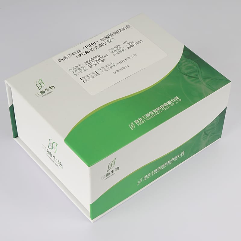


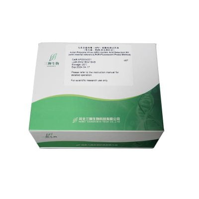
Item No: AP2409002 Specification: 50T/100T Storage: -20℃
Product introduction:
Pigeon Herpesvirus (PiHV) is a viral infectious disease primarily affecting young pigeons caused by Pigeon Herpesvirus Type 1 (PiHV1). It is an important pathogen leading to pigeon squab disease syndrome in young pigeons. Clinically, it is characterized by symptoms such as nasal and conjunctival inflammation. Infected pigeons may also exhibit depression, decreased appetite, diarrhea, vomiting, and neurological symptoms. Therefore, early, rapid, and convenient detection of PiHV is crucial for diagnosis, slowing the spread of the disease, and preventing complications. This test kit is designed based on the conserved sequences in the PiHV gene, with optimized conditions, enabling accurate fluorescent quantitative PCR testing of oral, cloacal swabs, blood, and other tissue samples from birds. This allows for a quick determination of whether the host is infected with PiHV.
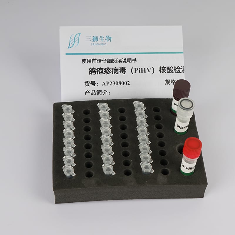
Product contents:
| Reagent components | AN2409002-02 (50T) | AN2409002-03 (100T) |
| PiHV Testing Reagent | 1150μL | 1150µL× 2 |
| PiHV Positive Control | 100μL | 100μL |
| Negative Control | 100μL | 100μL |
Storage conditions:
Store at -20℃, the shelf life is at least 12 months.
Experimental operation:
- Sample Preparation (Sample Preparation Area)
1.1 Sample Requirements:
-Oral/Clocal Swab: Use a sterile cotton swab to insert into the bird’s throat or cloaca, rotating three times. Remove it and place it in a centrifuge tube, cutting off the exposed part, then securely cap the tube and label it. Samples that can be tested within 24 hours can be stored at 2-8°C; samples that cannot be tested promptly should be stored at -20°C.
-Whole Blood: Use EDTA as an anticoagulant; do not use heparin. Fresh whole blood is required, or it can be stored at 2-8°C for no more than 7 days, or at -20°C for no more than 3 months. Repeated freeze-thaw cycles should be avoided as much as possible.
-Tissue: Take fresh liver tissue not exceeding 100 mg, and prepare a tissue homogenate using 200-500μL of sterile water for subsequent sample preparation.
1.2 Sample Preparation:
Refer to Sanshibio’s “Animal Genome DNA Rapid Extraction Kit (for PCR Analysis) Manual” (Catalog No.: DP202), or other nucleic acid extraction kits/methods that meet relevant requirements, to extract nucleic acids from processed samples. The extracted nucleic acid samples should be placed in an ice box and tested as soon as possible; they can be stored at 4°C for no more than 7 days or at -20°C for no more than 6 months.
- Prepare the Reaction System (Sample Addition Area)
2.1 Take out each component of the kit, completely thaw at room temperature, and centrifuge for 10 seconds to collect liquid from the tube walls and caps at the bottom. Prepare the reaction system according to the number of samples (n+2) (i.e., n test samples + 1 positive control + 1 negative control). Specifically, prepare (n+2) PCR tubes, dispensing 23μL of the PiHV Testing Reagent into each tube, and keep them in an ice box for later use.
2.2 Then, sequentially add 2μL of the Negative Control, extracted sample nucleic acid, and PiHV Positive Control to the testing reagent. Cap the tubes securely and record the information; the total volume for each reaction should be 25μL. Mix thoroughly, centrifuge for 10 seconds, and conduct the amplification experiment in the PCR instrument.
- PCR Amplification (Amplification and Product Analysis Area)
50°C UNG enzyme decontamination for 2 minutes; 95°C pre-denaturation for 2 minutes; 95°C denaturation for 10 seconds, followed by 60°C annealing and extension for 35 seconds, for 40 cycles, collecting fluorescence signals at 60°C. Select the fluorescence channel FAM (if using ABI series or similar real-time quantitative PCR instruments, contact the manufacturer in advance if needed or add ROX calibration dye yourself; otherwise, follow the normal procedure).
- Result Determination
4.1 Conditions for Validating Experimental Results:
Positive control FAM channel Ct value < 28 and exhibits an “S” shaped amplification curve; negative control has no Ct value or Ct value ≥ 38 and no “S” shaped amplification curve, indicating valid results; otherwise, repeat the experiment. If retesting remains invalid, please contact the manufacturer’s technical personnel.
4.2 Sample Result Determination:
Ct value ≤ 33 and exhibits an “S” shaped amplification curve, judged as PiHV positive;
Ct value 33 < Ct < 38 is considered suspicious; retesting is recommended (nucleic acid extraction may be substandard or there are strong inhibitory contaminants (e.g., residues from disinfectants, anticoagulants) inhibiting amplification; suggest reprocessing the sample for nucleic acid extraction and amplification; first rule out false positive results caused by aerosol contamination before re-extracting nucleic acids and retesting). If aerosol contamination is ruled out and the FAM channel retesting Ct value < 33 with a clear amplification curve, it is determined as PiHV positive; otherwise, it is determined as negative;
No Ct value or Ct value ≥ 38 and no “S” shaped amplification curve is determined as PiHV negative.
Precautions:
- To prevent contamination, the experiment should be strictly partitioned, and physical isolation between partitions is preferred to avoid cross-contamination due to human factors. Wear lab coats and latex gloves during the experiment, use tools independently in different areas, change gloves and lab coats when needed. After PCR, do not open the lid immediately; open it after sufficient cooling to minimize aerosol contamination.
- Thaw the reagents completely before use, but avoid repeated freeze-thaw cycles. The PCR reaction tubes provided in the kit are 0.2mL; if replacement is needed, transfer after complete thawing. Strictly follow the instructions for reagent preparation and sample addition. Workstations, centrifuges, pipettes, and other instruments should be disinfected regularly with chlorine-containing disinfectants, alcohol, nucleic acid decontamination agents, or UV light.
- A negative result does not necessarily mean the host is not infected with the virus. Poor sample quality, low virus load, or the presence of strong inhibitory substances (such as alcohol, disinfectants, anticoagulants, etc.) may result in unsuccessful nucleic acid extraction (detection) and a “negative” result. Follow the result interpretation criteria, and when there are suspicious results, first exclude aerosol contamination. If there are other questions, contact the manufacturer’s technical personnel.
- This product is for one-time use only. The components in the reagent are sensitive to natural light; avoid light exposure during packaging and storage. After packaging, do not repeatedly freeze-thaw. For scientific research use only; not for clinical diagnosis or other purposes.
Reviews
There are no reviews yet.